Arteries In Neck Diagram / Pin on EMT / In the following diagrams, the anatomy is drawn with and without the labels.. This diagram of the human heart shows all the major vessels, and arrows indicate the direction of flow through the heart. The left common carotid artery and left subclavian artery arising directly from the arch of the aorta to supply similar territories on the left side of the body. The first branch of the thyrocervical trunk is the inferior thyroid artery. Neck diagram of muscles, arteries, and skeleton. The right and left subclavian arteries give rise to the thyrocervical trunk.
The right and left subclavian arteries give rise to the thyrocervical trunk. The axillary artery is the main artery of the upper limb. We go into great detail on the flow of. It is divided into two portions. Neck dissection removes potential or proven metastases to cervical lymph nodes.
Anatomy And Physiology Coloring Pages Free at GetColorings ... from getcolorings.com In the following diagrams, the anatomy is drawn with and without the labels. The left common carotid artery and left subclavian artery arising directly from the arch of the aorta to supply similar territories on the left side of the body. The first branch of the thyrocervical trunk is the inferior thyroid artery. If one of them is narrowed or blocked, it can lead to a stroke. It runs from the heart down the length of the chest and abdomen. Note the feathery network of blood vessels in the left and right lungs (next to the heart). The brachiocephalic artery left common carotid artery and left subclavian artery. The right and left subclavian arteries give rise to the thyrocervical trunk.
The brachiocephalic artery left common carotid artery and left subclavian artery.
The brachiocephalic artery left common carotid artery and left subclavian artery. It passes under the neck of pancreas and supplies blood to the head and. Related online courses on physioplus. An artery is an elastic blood vessel that transports blood away from the heart. The right and left subclavian arteries give rise to the thyrocervical trunk. This section of the website will explain large and minute details of arterial anatomy of neck. The carotid artery pulse can be felt by pushing lateral to the upper border of the thyroid cartilage just under the anterior edge of the sternomastoid muscle. When you need a screening test—and when you don't. Illustration of the arterial system in the human body, shown in a standing figure. It is attached to the left ventricle of the heart you can feel your pulse in an artery such as the carotid artery in the neck or the radial artery in the hardening of the arteries is the common term for atherosclerosis and peripheral arterial disease. Ploaded with beautifully illustrated diagrams clearly and concisely labeled for easy identification. It runs from the heart down the length of the chest and abdomen. Posted by cassidy smith on 9 may 2018, 11:14 am.
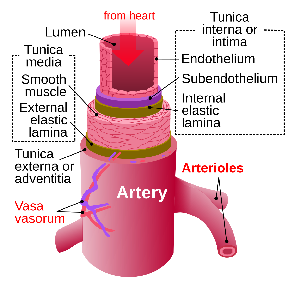
Related online courses on physioplus. Printable neck diagrams to help you learn more about the system that makes up our neck. We go into great detail on the flow of. It is bound laterally by the carotid arteries, superiorly by the hyoid bone, and inferiorly by. Smaller arteries are more muscular in the structure of their walls.
Upper Major Systemic Arteries | Human anatomy and ... from i.pinimg.com It is divided into two portions. They are the carotid arteries, and they carry blood to the brain. The first branch of the thyrocervical trunk is the inferior thyroid artery. Ninja nerds!join us in this video where we discuss the blood circulation of the head and neck using a flow chart. The descending aorta is the largest artery in the body; Smartdraw includes 1000s of professional healthcare and anatomy head & neck lateral view of the head with arteries of the head and neck shown in relation to underlying skeletal structures. Posted by cassidy smith on 9 may 2018, 11:14 am. The right and left subclavian arteries give rise to the thyrocervical trunk.
There are two large arteries in the neck, one on each side.
There are two large arteries in the neck, one on each side. Printable neck diagrams to help you learn more about the system that makes up our neck. In the following diagrams, the anatomy is drawn with and without the labels. Bodytomy provides a labeled celiac artery diagram to help you understand the location, anatomy, and function of this artery. Instant anatomy is a specialised web site for you to learn all about human anatomy of the body with diagrams, podcasts and revision questions. The main artery of the systemic circulation is the aorta. If one of them is narrowed or blocked, it can lead to a stroke. The left common carotid artery and left subclavian artery arising directly from the arch of the aorta to supply similar territories on the left side of the body. The neck diagram above shows you the structure and anatomy of the neck. From this trunk, several vessels arise, which go on to supply the neck. In the neck, the following diagram points out the major landmarks of the neck. It passes under the neck of pancreas and supplies blood to the head and. It is divided into two portions.
Thanks to easy access, the artery is useful in clinical procedures such as coronary angiography. Neck dissection removes potential or proven metastases to cervical lymph nodes. It passes under the neck of pancreas and supplies blood to the head and. It lies anterior to ica and is the chief arterial supply to structures in front of neck and face. The carotid artery pulse can be felt by pushing lateral to the upper border of the thyroid cartilage just under the anterior edge of the sternomastoid muscle.
Artery - Wikipedia from upload.wikimedia.org This diagram of the human heart shows all the major vessels, and arrows indicate the direction of flow through the heart. Deep lingual artery) is the terminal portion of the lingual artery; Start studying arteries head and neck. It begins at the outer border of the first rib as the continuation of subclavian artery and ends by becoming brachial artery at the lower. When you need a screening test—and when you don't. The main artery of the systemic circulation is the aorta. Common carotid, external carotid (and branches except maxillary, superficial temporal and posterior auricular), internal carotid artery (and sinus) veins: It passes under the neck of pancreas and supplies blood to the head and.
The first branch of the thyrocervical trunk is the inferior thyroid artery.
Printable neck diagrams to help you learn more about the system that makes up our neck. The internal carotid artery is located in the inner side of the neck in contrast to the external carotid artery. Whiplash associated disorders and neck rehabilitation online course: Illustration of the arterial system in the human body, shown in a standing figure. Deep lingual artery) is the terminal portion of the lingual artery; Instant anatomy is a specialised web site for you to learn all about human anatomy of the body with diagrams, podcasts and revision questions. Related online courses on physioplus. The neck diagram above shows you the structure and anatomy of the neck. The first branch of the thyrocervical trunk is the inferior thyroid artery. Master the triangles of the neck with our study unit which includes video tutorials, quizzes, labeled diagrams, and articles arteries: Ploaded with beautifully illustrated diagrams clearly and concisely labeled for easy identification. They are the carotid arteries, and they carry blood to the brain. When you need a screening test—and when you don't.
The first branch of the thyrocervical trunk is the inferior thyroid artery arteries in neck. The external carotid artery supplies the areas of the head and neck external to the start studying arteries of head and neck.
Berbagi :
Posting Komentar
untuk "Arteries In Neck Diagram / Pin on EMT / In the following diagrams, the anatomy is drawn with and without the labels."


Posting Komentar untuk "Arteries In Neck Diagram / Pin on EMT / In the following diagrams, the anatomy is drawn with and without the labels."