Shoulder Muscles Diagram Posterior - Handcuff Muscles Brooklyn Reflexology : Anterior graphic of the shoulder.. These muscles form the outer shape of the shoulder and underarm. The shoulder consists of three joints: You will hinder your progress if you overwork your shoulder muscles. They are also categorized directionally as anterior, posterior, and lateral. With cross body stretching, people will often why is this relevant?
General anatomy and the musculoskeletal system: Learn their origins/insertions, functions & exercises. This muscle diagram is interactive: Posterior part of the deltoid: (from schuenke m, schulte e.
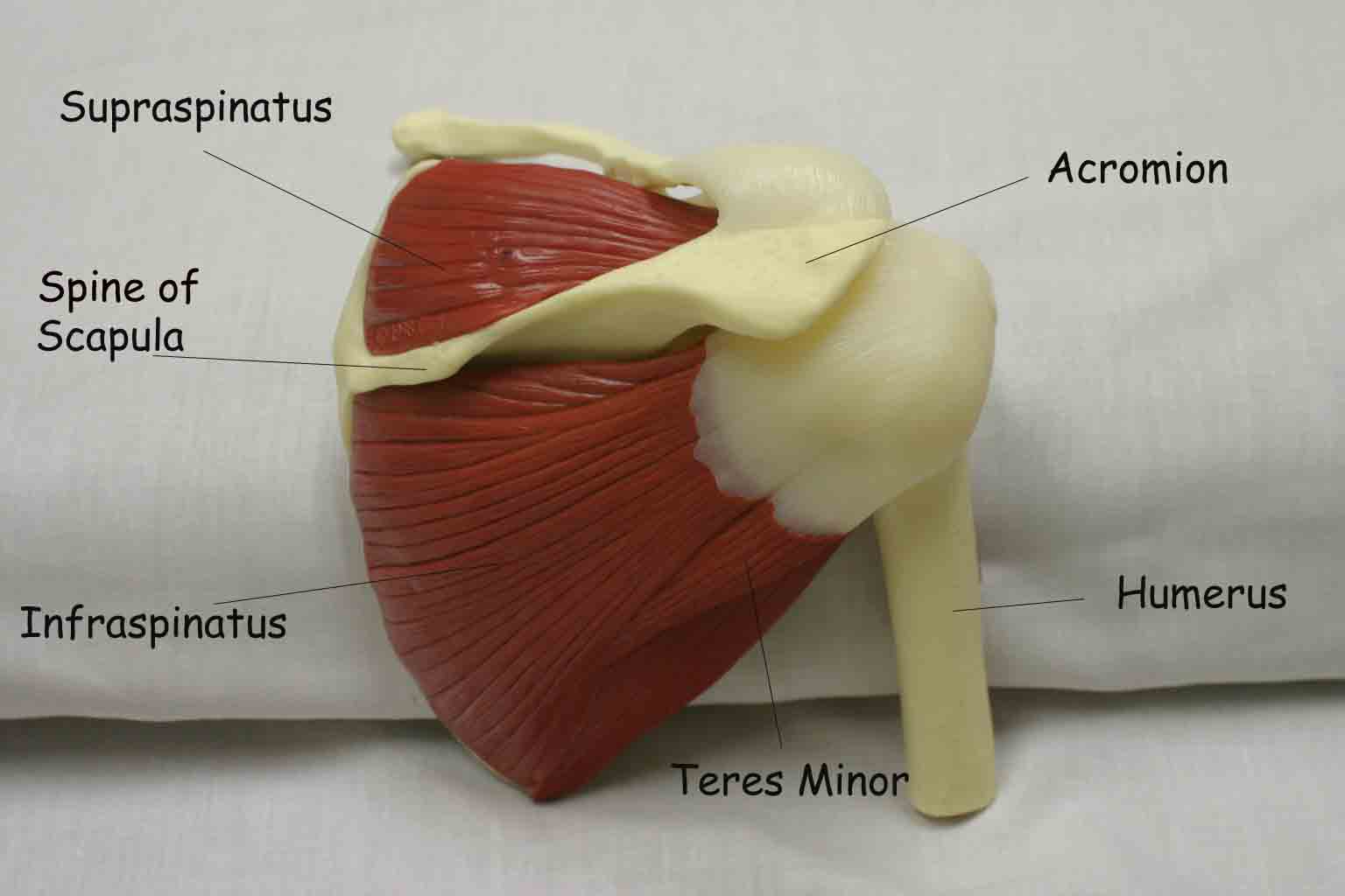
Anatomy Of The Upper Limb Shoulder Muscles Posterior View Youtube from i.ytimg.com Nine muscles cross the shoulder joint. The posterior muscles of the shoulder: Click on the name of a muscle for a page about that muscle (works for most labels). Want to learn more about it? Neck and shoulder muscles diagram muscles of neck anterior view dental hygiene pinterest anatomy. 7.16 posterior muscles of the shoulder and arm. Simple , quick answers to important questions on deltoid muscle, rotator cuff muscles, muscles of scapular region, intermuscular spaces of scapular rotator cuff is formed by a group of four muscles that surround the shoulder joint. They are shown in the image below.
Shoulder muscles and shoulder tendons.
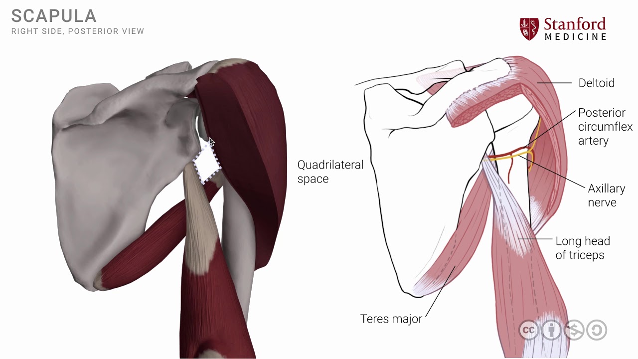
Summary of the structure of the posterior shoulder muscles. 7.16 posterior muscles of the shoulder and arm. Anterior graphic of the shoulder. Posterior band of the ighl. Unlike in other joints, the shoulder's dynamic stability and control are provided primarily by muscles (the rotator cuff in particular) rather. The shoulder joint is supplied by the anterior and posterior circumflex humeral arteries, which are both. The human shoulder is made up of three bones: The clavicle (collarbone), the scapula (shoulder blade), and the humerus (upper arm bone) as well as associated muscles, ligaments and tendons. For baseball pitchers, the teres minor demonstrates the highest level of emg activity of all the shoulder muscles during the. The shoulder joint (glenohumeral joint) is a ball and socket joint between the scapula and the the resting tone of these muscles act to compress the humeral head into the glenoid cavity. With seizure activity, the internal rotator muscles (teres major. Posterior shoulder mobility deficits often lead to limitations in shoulder internal rotation and horizontal adduction. Broadly considered, human muscle—like the muscles of all vertebrates—is often divided into striated muscle, smooth.
Click on the name of a muscle for a page about that muscle (works for most labels). Right posterior basal segmental bronchus. The shoulder muscles include skeletal muscles that are attached to the head of the humerus which performs various direct and indirect functions of the shoulder joints. Extends and laterally rotates the arm. Anterior graphic of the shoulder.
Uc San Diego S Practical Guide To Clinical Medicine from meded.ucsd.edu With seizure activity, the internal rotator muscles (teres major. Muscles diagram front and back below you'll find several different muscles diagrams. The glenohumeral joint (commonly referred to as shoulder joint), the sternoclavicular joint, and the acromioclavicular joint. Want to learn more about it? The shoulder muscles include skeletal muscles that are attached to the head of the humerus which performs various direct and indirect functions of the shoulder joints. Shoulder muscles and shoulder tendons. Unlike in other joints, the shoulder's dynamic stability and control are provided primarily by muscles (the rotator cuff in particular) rather. Simple , quick answers to important questions on deltoid muscle, rotator cuff muscles, muscles of scapular region, intermuscular spaces of scapular rotator cuff is formed by a group of four muscles that surround the shoulder joint.
Want to learn more about it?
Posterior muscles in the body. Right posterior basal segmental bronchus. The anterior, lateral and posterior deltoid heads. The shoulder has about eight muscles that attach to the scapula, humerus, and clavicle. Muscles of the shoulder can be divided into two strata: Infraspinatus and teres minor tendon. Summary of the structure of the posterior shoulder muscles. Learn vocabulary, terms and more with flashcards, games and other study tools. This muscle diagram is interactive: Posterior part of the deltoid: The muscles (and associated muscle tissues) labelled in the posterior muscles diagram shown above are listed in bold the following table by part. Important muscular spaces of shoulder. (from schuenke m, schulte e.
Medical illustration of the shoulder's muscles : Posterior part of the deltoid: They are also categorized directionally as anterior, posterior, and lateral. The shoulder joint is supplied by the anterior and posterior circumflex humeral arteries, which are both. Posterior muscles in the body.
Shoulder Axilla And Brachial Plexus Amboss from media-us.amboss.com Anteriorly and posteriorly the muscles attach on each side of the depressions (groove and sulcus). Posterior humerus, superior to the radial groove medial head: Infraspinatus and teres minor tendon. Medical illustration of the shoulder's muscles : The shoulder muscles include skeletal muscles that are attached to the head of the humerus which performs various direct and indirect functions of the shoulder joints. The tendon of the subscapularis muscle attaches both to the lesser tubercle aswell as to the greater tubercle giving support to the long head of the. 7.24 linear diagram of brachial plexus and branches. Superficial layer with deltoid, trapezius, pectoralis.
General anatomy and the musculoskeletal system:
The anterior deltoid, the lateral deltoid, and the posterior deltoid. Important muscular spaces of shoulder. Anterior graphic of the shoulder. There are three main muscles in your shoulder: With cross body stretching, people will often why is this relevant? Related online courses on physioplus. Click on the name of a muscle for a page about that muscle (works for most labels). General anatomy and the musculoskeletal system: Posterior shoulder mobility deficits often lead to limitations in shoulder internal rotation and horizontal adduction. The shoulder has about eight muscles that attach to the scapula, humerus, and clavicle. (from schuenke m, schulte e. With seizure activity, the internal rotator muscles (teres major. Anterior part of the deltoid:
Simple , quick answers to important questions on deltoid muscle, rotator cuff muscles, muscles of scapular region, intermuscular spaces of scapular rotator cuff is formed by a group of four muscles that surround the shoulder joint shoulder muscles diagram. Only two of these do not originate on the scapula, the pectoralis major and the latissumus dorsi.
Berbagi :
Posting Komentar
untuk "Shoulder Muscles Diagram Posterior - Handcuff Muscles Brooklyn Reflexology : Anterior graphic of the shoulder."


Posting Komentar untuk "Shoulder Muscles Diagram Posterior - Handcuff Muscles Brooklyn Reflexology : Anterior graphic of the shoulder."